Prenatal Care & Testing
Molar Pregnancy: What It Is, Symptoms and Treatment
A molar pregnancy is a rare form of gestational trophoblastic disease that looks like a pregnancy on a test but isn't. Here is what the evidence says about diagnosis, treatment, and trying again.
Clinically reviewed · June 2026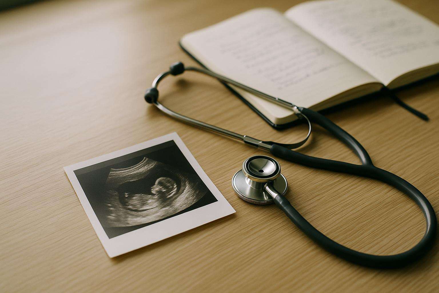
A molar pregnancy is a rare, non-viable growth of abnormal placental tissue—complete or partial—that requires surgical removal and careful hCG monitoring afterward. It is not caused by anything you did, and most women go on to have healthy pregnancies once the monitoring period is complete.
Receiving a molar pregnancy diagnosis is disorienting in a particular way. You tested positive. You may have had symptoms that felt like a real pregnancy. And then an ultrasound or bloodwork revealed something entirely different. Understanding exactly what a molar pregnancy is—the biology, the two types, the treatment, and the path forward—is the most useful thing you can have right now alongside your care team.
A note before we begin: this article provides general educational information grounded in current medical evidence. It is not a substitute for evaluation and individualized care from a qualified OB-GYN or gynecologic oncologist. If you have been diagnosed with or suspect a molar pregnancy, please contact your provider promptly.
What Exactly Is a Molar Pregnancy?
A molar pregnancy—its clinical name is hydatidiform mole—belongs to a family of conditions called gestational trophoblastic disease (GTD). GTD encompasses a spectrum of abnormal growths that arise from the trophoblast, the layer of cells that normally forms the placenta. In a molar pregnancy specifically, an error at or immediately after fertilization causes placental cells to proliferate in an uncontrolled way, forming clusters of fluid-filled sacs sometimes described as resembling a bunch of grapes on ultrasound.
The condition produces a positive pregnancy test because the abnormal trophoblastic cells secrete human chorionic gonadotropin (hCG)—the same hormone that all pregnancy tests detect. In many molar pregnancies, hCG is produced at levels far higher than in a normal pregnancy, sometimes reaching hundreds of thousands of mIU/mL. According to a 2024 review of hCG dynamics in early gestational events published in Obstetrics and Gynecology International, hCG in a normal singleton pregnancy peaks at roughly 50,000–100,000 mIU/mL around 8–10 weeks and then declines. Molar pregnancies can far exceed this range, and the extreme elevation itself can sometimes produce a paradoxically faint or negative result on a standard home test through a mechanism known as the hook effect.
A molar pregnancy cannot develop into a viable baby. Both types—complete and partial—require removal and monitoring.
Complete vs. Partial Molar Pregnancy: What Makes Them Different?
The two types differ in their genetic origin, appearance, and risk profile. Distinguishing them matters clinically because it shapes follow-up duration and the likelihood of requiring treatment beyond surgical evacuation.
Complete molar pregnancy occurs when a sperm fertilizes an egg that has lost its genetic material—essentially an empty egg. The resulting tissue is entirely paternal in origin, carrying a duplicated set of chromosomes (typically 46,XX or 46,XY) but no maternal DNA. No embryo or fetal tissue forms. The uterus fills with the characteristic grape-like molar mass, and hCG levels rise markedly—often into the very high range. Complete moles carry approximately a 15–20% risk of developing persistent GTD after evacuation, meaning the tissue does not resolve fully on its own and chemotherapy becomes necessary.
Partial molar pregnancy occurs when two sperm fertilize a single egg simultaneously, creating a triploid conception with 69 chromosomes instead of the normal 46. Some embryonic or fetal tissue may be present—occasionally enough to be visible on early ultrasound—but it is genetically abnormal and cannot survive. Partial moles produce lower hCG levels than complete moles, and the risk of persistent GTD is substantially lower at roughly 0.5–5%.
The definitive distinction between a complete and partial mole—and between either mole type and other causes of first-trimester loss—is made by pathological examination of the evacuated tissue, not by ultrasound or symptom pattern alone. Early moles in particular can look non-specific on imaging, which means histology is essential.
Many molar pregnancies are now identified earlier than they were in prior decades, when the classic grape-cluster appearance on ultrasound was the primary diagnostic cue. First-trimester ultrasound at 8–10 weeks often reveals a snowstorm pattern or other abnormality before symptoms become prominent. Earlier detection has improved outcomes by enabling earlier treatment.
What Are the Symptoms of a Molar Pregnancy?
Because molar pregnancies produce hCG, many early symptoms are indistinguishable from a normal first trimester. The features that should prompt further evaluation are:
- Vaginal bleeding in the first trimester. This is the most common presenting symptom, occurring in the majority of diagnosed cases. Unlike the light spotting that can accompany implantation, bleeding associated with a molar pregnancy can be heavier and is often accompanied by the passage of small, fluid-filled sacs of tissue.
- Severe nausea and vomiting. The markedly elevated hCG levels in complete moles can produce hyperemesis—vomiting severe enough to interfere with daily function or require medical management—more commonly and more intensely than in typical pregnancy.
- Uterus measuring large for dates. The rapid growth of molar tissue can cause the uterus to expand faster than expected, which a provider notices during a pelvic examination.
- Preeclampsia before 20 weeks. Hypertension with proteinuria before 20 weeks of gestation is a red flag for molar pregnancy because preeclampsia almost never develops this early in a normal pregnancy.
- Signs of hyperthyroidism. Very high hCG levels can cross-activate thyroid-stimulating hormone receptors, occasionally producing palpitations, tremor, or other hyperthyroid symptoms in women with complete moles.
Partial moles are more often clinically silent in early pregnancy and are frequently discovered incidentally on a first-trimester ultrasound or when pathology is performed on tissue from what appeared to be a routine miscarriage.
How Is a Molar Pregnancy Diagnosed?
Diagnosis rests on a combination of serum beta-hCG measurement and pelvic ultrasound, confirmed by pathological analysis of evacuated tissue.
On ultrasound, a complete molar pregnancy in the second trimester classically shows a heterogeneous, echogenic intrauterine mass with multiple small cystic spaces—the so-called snowstorm appearance—with no identifiable fetal parts. First-trimester complete moles may appear less distinctive, sometimes resembling an incomplete or anembryonic pregnancy. A partial mole may show Swiss-cheese cystic changes within the placenta alongside an abnormal fetus or fetal pole with multiple structural anomalies.
Because the ultrasound picture can be ambiguous, especially early in the first trimester, serum beta-hCG is an important complementary tool. A level disproportionately high for gestational age—or one that is rising in a pattern inconsistent with normal pregnancy—strengthens suspicion. The hook effect documented in PubMed Central research on gestational trophoblastic disease means that a standard home urine test may paradoxically read negative or faintly positive when hCG is extremely elevated: the test's antibody system becomes saturated and cannot form the signal complex correctly. If clinical suspicion is present, a serum test—performed with serial dilution to avoid the hook effect—is essential.
Final diagnosis is confirmed when evacuated tissue is sent to pathology. Even a histologically straightforward case benefits from complete diagnosis to confirm the mole type and rule out more aggressive GTD variants such as invasive mole or, very rarely, choriocarcinoma.
How Is a Molar Pregnancy Treated?
Treatment has two sequential components: evacuation of the uterus and post-evacuation monitoring.
Surgical evacuation. For women who wish to preserve fertility, the standard approach is suction curettage (vacuum aspiration), performed under general or regional anesthesia. The procedure removes molar tissue from the uterine cavity. Oxytocin is often administered simultaneously to help the uterus contract and minimize blood loss. For women who have completed their family, hysterectomy is an alternative that eliminates the risk of local uterine recurrence, though it does not eliminate the need for subsequent hCG surveillance to detect distant disease.
Medical management (prostaglandins, mifepristone) is not recommended as primary treatment for molar pregnancy because it does not reliably clear all trophoblastic tissue and delays diagnosis through histological examination.
hCG surveillance after evacuation. After surgery, serial serum beta-hCG measurements are the cornerstone of follow-up. Levels should fall progressively toward zero after evacuation. Most centers measure hCG weekly until undetectable, then monthly for a defined surveillance period—typically six months for partial moles and twelve months for complete moles, though protocols vary by institution and risk profile.
During the monitoring period, reliable contraception (combined oral contraceptives are generally preferred and do not affect hCG clearance) is strongly recommended. A new pregnancy would raise hCG and make it impossible to distinguish normal early pregnancy from persistent or recurrent GTD without an established zero baseline.
Persistent GTD and chemotherapy. If hCG levels plateau or begin to rise during surveillance, persistent gestational trophoblastic disease is diagnosed. This occurs in roughly 15–20% of complete moles and 0.5–5% of partial moles. Persistent GTD does not mean cancer in the conventional sense—most cases are locally invasive moles—but it requires chemotherapy. Single-agent methotrexate is the standard first-line treatment for low-risk persistent GTD, with cure rates exceeding 95%. Actinomycin D is an alternative for methotrexate-resistant or -intolerant cases. High-risk disease (choriocarcinoma or placental site trophoblastic tumor) requires multi-agent regimens and specialist management from a gynecologic oncologist.
What Happens After a Molar Pregnancy — and When Can You Try Again?
One of the most important questions after a molar pregnancy diagnosis is: how long do I need to wait before trying to conceive again?
The answer depends on mole type and the protocol of the managing center. General guidance from clinical consensus:
- Partial molar pregnancy: Most centers recommend waiting until hCG has been undetectable for at least six months.
- Complete molar pregnancy: The standard recommendation is to wait until hCG has been undetectable for twelve months, because complete moles carry a higher risk of persistent GTD and a longer surveillance window is needed to confirm resolution.
The risk of a molar pregnancy recurring in a subsequent pregnancy is approximately 1–2%—modestly higher than the general population risk but not dramatically so. Reassuringly, once surveillance is complete and a new pregnancy begins, ACOG clinical guidance supports that prior molar pregnancy does not increase the risk of miscarriage, birth defects, or other complications in subsequent pregnancies. Early ultrasound and hCG monitoring in the next pregnancy are typically recommended so any concern can be identified promptly.
The emotional weight of a molar pregnancy is substantial and should not be minimized. You were pregnant, or at least expecting to be—and navigating an unexpected, non-viable diagnosis followed by surgery and months of monitoring is genuinely hard. Many women find it helpful to connect with support communities for pregnancy loss and with a perinatal counselor during the surveillance period. The waiting period, though frustrating, is purposeful—and research supports good outcomes on the other side of it for the vast majority of women who go through this experience.
Work closely with your OB-GYN or gynecologic oncologist throughout the monitoring phase. Keep every follow-up appointment, use the recommended contraception consistently, and contact your care team promptly if you have any symptoms—bleeding, pelvic pain, shortness of breath, or neurological changes—during surveillance. These are not signs to watch and wait on.
Frequently asked
What is a molar pregnancy?
A molar pregnancy—medically called a hydatidiform mole—is a type of gestational trophoblastic disease (GTD) in which abnormal trophoblastic tissue grows in the uterus instead of a normal pregnancy. Rather than a healthy embryo and placenta developing, an error at fertilization causes placental cells to multiply rapidly and form grape-like clusters of tissue. The condition tests positive for pregnancy because the abnormal tissue still produces human chorionic gonadotropin (hCG)—often at extremely high levels. Molar pregnancies cannot develop into a live birth. They require prompt medical removal and careful follow-up monitoring to ensure all abnormal tissue has resolved and, in rare cases, to watch for the development of persistent GTD or, very rarely, gestational trophoblastic neoplasia. This is general information, not medical advice. If you suspect a molar pregnancy, contact your provider immediately.
What is the difference between a complete and a partial molar pregnancy?
A complete molar pregnancy occurs when a sperm fertilizes an egg with no genetic material (an empty egg), resulting in tissue that is entirely paternal in origin. The uterus contains no fetal tissue—only the abnormal grape-like molar mass. Complete moles produce very high hCG levels and carry a higher risk (roughly 15–20%) of progressing to persistent GTD requiring further treatment.
A partial molar pregnancy occurs when two sperm fertilize a single egg, resulting in a triploid (69-chromosome) conception. Some fetal or embryonic tissue may be present alongside the molar tissue, but it cannot survive. Partial moles produce lower hCG levels and carry a lower risk (roughly 0.5–5%) of persistent GTD. Both types are diagnosed by pathological examination of tissue after evacuation, not by ultrasound or symptoms alone. Always discuss your specific diagnosis with your OB-GYN or gynecologic oncologist.
What are the symptoms of a molar pregnancy?
Molar pregnancy symptoms overlap significantly with normal early pregnancy, which is why most are not detected until an ultrasound or abnormal lab work raises concern. The most common warning signs include: vaginal bleeding in the first trimester, which occurs in the majority of cases; severe nausea and vomiting (hyperemesis) more intense than typical morning sickness; a uterus that measures larger than expected for gestational age; and the passage of grape-like tissue vaginally. Less commonly, women with complete moles may develop early-onset preeclampsia before 20 weeks—a timing that should always trigger further evaluation—or signs of hyperthyroidism driven by very high hCG levels cross-activating thyroid receptors. If you experience heavy bleeding, passage of tissue, or severe vomiting in early pregnancy, contact your provider the same day.
Why does a molar pregnancy cause a very high hCG level and what is the hook effect?
Molar pregnancies—particularly complete moles—produce abnormally large amounts of hCG because the rapidly proliferating trophoblastic cells act as an outsized hormonal factory. Where a normal singleton pregnancy reaches peak hCG of roughly 50,000–100,000 mIU/mL around 8–10 weeks, complete moles can produce hCG in the hundreds of thousands. This matters for home testing because of a phenomenon called the hook effect (prozone effect): when hCG levels are extremely high, they can overwhelm a standard test's antibody system and paradoxically produce a falsely negative or faintly positive result. As documented in research published on PubMed Central / NCBI, the hook effect is a recognized cause of false-negative home tests in gestational trophoblastic disease. A serum beta-hCG blood test, which uses serial dilution, is not subject to this limitation and is the appropriate test when molar pregnancy is suspected.
How is a molar pregnancy treated?
The primary treatment is surgical evacuation—most commonly suction curettage (vacuum aspiration) performed under general or regional anesthesia. The procedure removes all molar tissue from the uterus. For women who have completed their family, hysterectomy is an alternative that eliminates the risk of local uterine recurrence, though it does not eliminate the need for hCG monitoring.
After evacuation, serial serum beta-hCG measurements are taken weekly until levels reach an undetectable range, then monthly for six months to one year, depending on the mole type and the protocol of the treating center. If hCG levels plateau or rise during follow-up, persistent GTD is diagnosed and chemotherapy—most often single-agent methotrexate—is initiated. Persistent GTD is highly treatable, with cure rates exceeding 95% with appropriate management. Reliable contraception is recommended throughout the monitoring period to avoid confusion between rising hCG from a new pregnancy and a persisting mole.
When can I try to get pregnant again after a molar pregnancy?
Most specialists and professional guidance recommend waiting until hCG levels have been undetectable for 6–12 months, depending on mole type and institutional protocol. Complete moles typically require a full 12 months of follow-up before attempting conception; partial moles may allow a shorter monitoring window of 6 months, though practices vary. The waiting period is not arbitrary: a new pregnancy raises hCG and makes it impossible to distinguish recurrent or persistent GTD from normal early pregnancy without a baseline of zero. Once monitoring is complete and hCG has remained undetectable for the required window, the risk of recurrent molar pregnancy is low—approximately 1–2%—and subsequent pregnancies carry no greater risk of miscarriage, birth defects, or complications than in the general population according to ACOG clinical guidance. Discuss your individual timeline with your OB-GYN or gynecologic oncologist.