Trimester by Trimester
The 20-Week Anatomy Scan: What It Screens For and What It Costs
A system-by-system guide to the Level II ultrasound — what the sonographer checks, what the scan can and cannot detect, and how to navigate self-pay costs ranging from $75 to $1,200.
Clinically reviewed · June 2026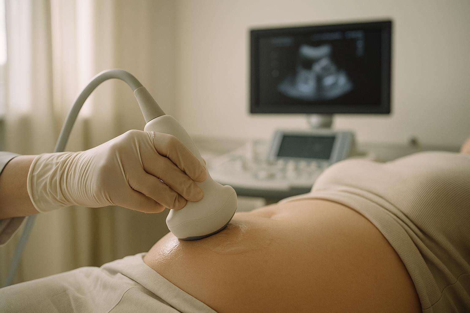
The 20-week anatomy scan is a 45-minute Level II ultrasound performed between 18 and 22 weeks. A sonographer systematically images your baby's brain, heart, spine, kidneys, limbs, placenta, and amniotic fluid. It screens for structural anomalies but cannot detect chromosomal conditions or guarantee a perfectly healthy outcome. Cost without insurance runs from $75 at community health clinics to $1,200 or more at hospital outpatient departments.
Somewhere around the halfway mark of your pregnancy, your provider will schedule what most families simply call "the 20-week scan." Its formal name — the Level II ultrasound or fetal anatomy survey — better captures what it actually is: the most thorough imaging evaluation of your baby that pregnancy medicine routinely performs. For many families it is also the appointment where fetal sex is first confirmed. This guide walks through every system the sonographer evaluates, what the scan realistically can and cannot tell you, and how to prepare for the cost if you are paying out of pocket or still working through your deductible.
What does the sonographer actually check during the anatomy scan?
The anatomy scan is conducted transabdominally — a sonographer applies conductive gel to your abdomen and moves a handheld transducer across the surface, converting reflected sound waves into real-time images on a screen. The session typically lasts 45 minutes or more, reflecting the sheer number of structures that must be measured and documented, according to Cleveland Clinic.
Here is a system-by-system overview of what the sonographer evaluates:
- Brain and neural tube. The cerebral ventricles, cerebellum, and spine are imaged to rule out neural tube defects such as spina bifida and anencephaly, and to check for hydrocephalus (fluid accumulation in the brain cavities).
- Face. The nose, lips, and palate are assessed to the extent that imaging allows; this is the screen for cleft lip (cleft palate is harder to visualize by ultrasound).
- Heart. A four-chamber view checks the basic cardiac architecture — size, position, and the relative proportions of atria and ventricles. The outflow tracts are also assessed when visualization permits. Congenital heart disease is the most common category of structural anomaly, and this portion of the scan receives particular attention.
- Spine. The vertebral column is imaged in its entirety for alignment and to screen for open neural tube defects along the back.
- Abdominal wall and organs. The sonographer confirms the abdominal wall is intact (ruling out gastroschisis and omphalocele), and checks that the stomach, intestines, liver, and diaphragm appear normal.
- Kidneys and bladder. Both kidneys are located and measured; the bladder should be visible and show normal filling. Absent or echogenic kidneys, dilated collecting systems, and cysts are noted for follow-up.
- Limbs. All four limbs — including hands and feet — are counted and measured. Femur and humerus lengths are recorded and compared to gestational age norms.
- Placenta and amniotic fluid. Placental location is documented to rule out placenta previa (a placenta covering the cervix). Amniotic fluid volume is estimated to check for oligohydramnios or polyhydramnios.
- Umbilical cord blood flow. Doppler assessment of cord flow confirms normal placental circulation.
If the baby is positioned appropriately and the family wishes to know, fetal sex is typically identified at this scan as well — though the sonographer will always note that no determination is 100% certain by ultrasound alone.
The anatomy scan is only as complete as your baby's cooperation. If the fetus is lying with its chest facing away from the transducer, or if the ribs create acoustic shadowing, the sonographer may not be able to obtain adequate cardiac views. You may be asked to walk, change position, or drink water and return — and in some cases a repeat scan is scheduled. This is normal, not a warning sign.
What can the anatomy scan miss — and what are its real limits?
Understanding what the anatomy scan cannot detect is just as important as knowing what it checks. The scan is a structural survey — it evaluates physical form, not chromosomal content, biochemistry, or functional neurological characteristics.
Congenital heart defects. Heart anomalies are the most common structural finding, and ironically the cardiac structures can be among the most difficult to fully evaluate. Sensitivity for congenital heart disease varies based on equipment quality, sonographer skill, and fetal position. Small ventricular septal defects and coarctation of the aorta, for instance, are frequently missed even under excellent conditions. A normal cardiac view at 20 weeks is reassuring but not definitive.
Chromosomal conditions. Trisomy 21 (Down syndrome), trisomy 18, and trisomy 13 may or may not produce structural findings visible at the anatomy scan. Some fetuses with chromosomal conditions have entirely normal-appearing anatomy on ultrasound. If chromosomal screening is a priority, non-invasive prenatal testing (NIPT) or diagnostic amniocentesis — not the anatomy scan — is the appropriate tool.
Late-emerging anomalies. Some kidney and urinary tract problems, certain brain abnormalities (such as agenesis of the corpus callosum), and some cardiac defects only become apparent later in gestation as structures mature. A follow-up growth scan in the third trimester may detect findings that were not present or not visible at 20 weeks.
Functional differences. The anatomy scan says nothing about cognitive development, autism spectrum disorder, behavioral characteristics, or hearing ability. These cannot be assessed by structural imaging at any gestational age.
When the anatomy scan identifies a finding — whether an isolated soft marker like a choroid plexus cyst, or a structural anomaly — the standard next step is referral to maternal-fetal medicine (MFM) for a targeted ultrasound and additional workup. That referral is the system working exactly as designed, not a cause for alarm in itself.
How much does the anatomy scan cost, and what should you ask before you go?
Self-pay cost for the anatomy scan varies substantially by setting, geography, and whether 3D or 4D imaging is added. Based on data from Beautiful Beginnings Ultrasound and NextGen Diagnostic Imaging, the typical ranges by facility type are:
- Community health clinics: $75–$150
- Independent imaging centers: $150–$800
- Hospital outpatient departments: $500–$1,200 or more
The hospital premium is not arbitrary. Hospitals routinely apply a facility fee on top of the radiologist or MFM specialist's professional fee — two separate line items for one appointment. Urban markets carry higher rates than rural areas, and 3D or 4D add-ons increase the final invoice further.
If you have insurance: Most major commercial plans cover the anatomy scan as a medically necessary prenatal service — it is a standard-of-care procedure. But coverage does not mean zero cost. You may still owe a deductible balance, a copay, and coinsurance. If the scan falls early in the year before your deductible resets, your out-of-pocket share can approach the self-pay rate. Before you go, call member services and ask three questions: Is the anatomy scan covered? Is my provider in-network? What is my remaining deductible balance?
If you are paying out of pocket: Ask your ordering provider for the relevant CPT code in advance — typically 76805 for a standard second-trimester survey. Call at least two or three imaging centers with that code and ask for their self-pay rate. Differences of several hundred dollars between facilities in the same metro area are common. Independent imaging centers almost always charge significantly less than hospital outpatient departments for an identical scan.
This article is general information, not medical advice. If you have questions about your anatomy scan results or what findings mean for your specific pregnancy, talk with your OB-GYN, CNM, or maternal-fetal medicine specialist.
Frequently asked
What exactly does the 20-week anatomy scan look at?
The anatomy scan, formally called the Level II ultrasound, is a systematic survey of your baby's major organ systems. According to the Cleveland Clinic, the sonographer evaluates the fetal brain and neural tube (checking for neural tube defects and hydrocephalus), the face (screening for cleft lip), the four-chamber heart (assessing for congenital cardiac defects), the spine alignment, the abdominal wall, both kidneys and bladder, the stomach, the diaphragm, and all four limbs including hands and feet. The sonographer also documents placental location to rule out placenta previa, measures amniotic fluid volume, and assesses umbilical cord blood flow. If the baby is positioned appropriately and you wish to know, fetal sex can typically be identified at this scan as well.
How long does the anatomy scan take and does it hurt?
The anatomy scan is a non-invasive, non-ionizing procedure — there is no radiation and no discomfort beyond the cold gel applied to the abdomen. The sonographer moves a handheld transducer across your skin, converting reflected sound waves into real-time images on a monitor. Cleveland Clinic notes that the session typically lasts 45 minutes or more — longer than most people expect. The extra time reflects the thoroughness of the survey: every structure is measured and documented systematically. If the baby is lying in an awkward position, you may be asked to walk around or lie on your side to encourage repositioning. Bring water, use the restroom beforehand rather than arriving with a full bladder (a full bladder is generally not required at 20 weeks as it was for earlier scans), and plan for the appointment to run up to an hour.
What can the anatomy scan miss?
The anatomy scan is thorough but not infallible. According to Cleveland Clinic, the scan's sensitivity for congenital heart defects — the most common structural anomaly — varies depending on equipment quality, the sonographer's experience, and fetal positioning. When the baby's chest faces away from the transducer, adequate cardiac views may be impossible. Minor defects, including small ventricular septal defects, can be missed even under ideal conditions. The scan also has limited ability to detect chromosomal conditions (that requires NIPT or amniocentesis), some kidney and urinary tract anomalies that appear only later in gestation, and functional neurological differences such as autism spectrum disorder. Abnormal findings prompt referral to maternal-fetal medicine (MFM) for targeted imaging and workup — exactly the system working as designed. A normal anatomy scan is genuinely reassuring, not a guarantee.
How much does the anatomy scan cost without insurance?
Self-pay costs vary significantly by setting and geography. Beautiful Beginnings Ultrasound and NextGen Diagnostic Imaging document the following typical ranges: community health clinics, $75–$150; independent imaging centers, $150–$800; hospital outpatient departments, $500–$1,200 or more. The hospital premium exists because hospitals typically layer a facility fee on top of the radiologist or MFM professional fee — two separate charges on one visit. Urban markets with higher costs of living carry higher rates than rural areas, and 3D or 4D add-ons increase the price further. If you are paying out of pocket, ask the ordering provider for the relevant CPT code in advance and call multiple imaging centers to compare self-pay rates before scheduling — differences can exceed several hundred dollars for an identical procedure.
Does insurance cover the anatomy scan, and what will I actually owe?
Most major commercial insurance plans cover the anatomy scan as a medically necessary prenatal service. However, coverage does not mean zero cost. As Beautiful Beginnings Ultrasound notes, depending on your plan, you may still owe a deductible, a copay, and/or coinsurance. If the scan happens early in the calendar year before your deductible has been met, your out-of-pocket share can be substantial — sometimes matching or approaching the self-pay rate. Before the appointment, call the member services number on your insurance card and ask: (1) Is the 20-week anatomy scan covered? (2) Is my provider in-network? (3) What is my current deductible balance? Also confirm whether the reading radiologist or MFM specialist bills under a different tax ID than the facility, as each may apply separately toward your deductible.
When is the anatomy scan scheduled, and can it be done earlier or later than 20 weeks?
The anatomy scan is performed between 18 and 22 weeks of gestation — a window that allows enough fetal growth for structures to be clearly visible while leaving time for follow-up if needed. As Cleveland Clinic explains, most practices aim for 19–20 weeks as a practical midpoint. Scheduling before 18 weeks risks incomplete visualization of still-forming structures; scheduling after 22 weeks compresses the time available for follow-up workup. If your scan falls at 18 weeks and the baby is poorly positioned, your provider may order a repeat targeted scan — particularly if risk factors are present such as a prior anomaly, abnormal NIPT result, or elevated nuchal translucency. A perfectly normal anatomy scan at 22 weeks is just as reassuring as one at 20: the exact week within the window matters less than ensuring the full survey is completed.